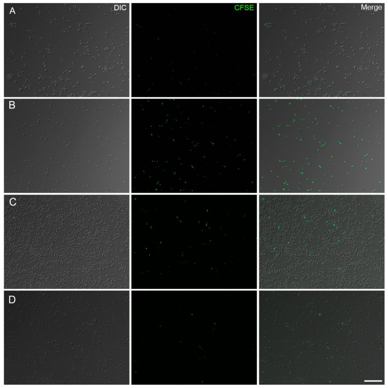
Figure 3.
Fluorescence images of CFSE stained A. baumannii after treatment with melittin (142 µg/mL) for 2 h at 37 °C. The proliferation of untreated bacteria (A) and bacteriostatic effect of melittin against ATCC strain (B). A. baumannii PDR strain 100 images of CFSE-labeled bacteria from untreated (C) and melittin treated cells (D). DIC: Differential interference contrast. Bar = 20 μm.

