Figure 1:
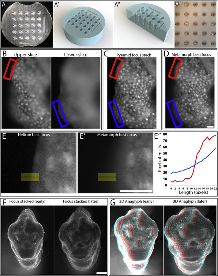
(A) Focus stacked image of mold used to produce tapered agarose wells (A’,A”) for long-term ‘anterior-up’ imaging. These wells are designed to orient the embryo with its face pointing towards the objective throughout its early development (A”’). (B) 2 separate focal planes of a single 3D sample with sample boundary being highlighted with red and blue boxes. (C) Helicon Pyramid focus and (D) Metamorph best focus stacks of the image in (B). (E’-E”’) Plotting pixel intensity over the same linear ROI of an image stacked with Helicon (E) or Metamorph (E’) shows that Helicon focus stacking produces a much sharper boundary (E”). (F) 2D focus stacks and (G) 3D anaglyphs produced with Helicon Focus using Mitotracker to produce contrast. Scale bar = 50 μm (B-E), 250 μm (F).

