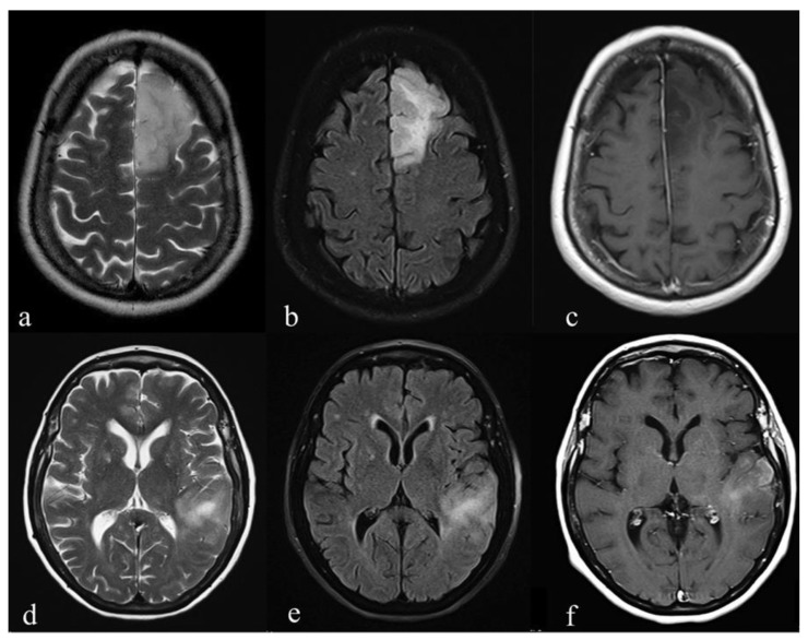
Figure 3.
Pre-surgical conventional MR images of two patients with Grade III astrocytoma: (a–c) T2-weighted, fluid-attenuated inversion recovery images (FLAIR) and T1-weighted post-gadolinium images of an IDH-mutant (Mut) tumor show a lesion on the left frontal lobe. The borders are well-defined on T2-weighted (a) and FLAIR (b) images. No areas of T1-shortening are evident after IV contrast medium administration; (d–f) T2-weighted, FLAIR and T1-weighted post-gadolinium of an IDH-wild type (Wt) tumor show a hyperintense T2-weighted (d) and FLAIR (e) lesion on the left temporal lobe. Ill-defined borders (d,e) and a blurred contrast enhancement are detected (f). Scale bar: 5 cm.

