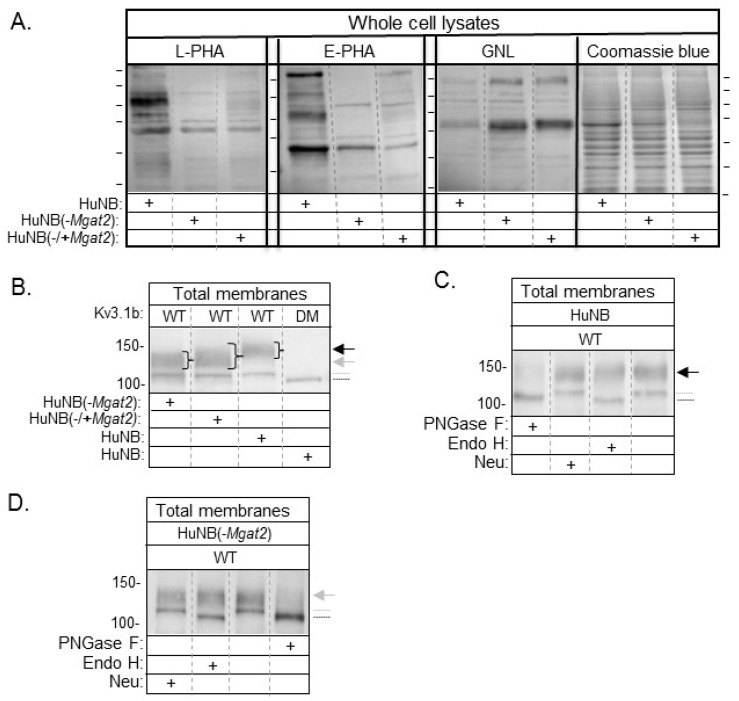
Figure 2.
Identification of reduced levels of complex type N-glycans in NB cells with MGAT2 silenced. Lectin blots of whole cell lysates from HuNB and HuNB(-MGAT2) cell lines, along with the later cell line transiently transfected with MGAT2 to create the rescued cell line, called NB_1(-/+MGAT2) (A). Proteins separated on membranes were probed with L-PHA, E-PHA, and GNL, as indicated. Coomassie blue-stained SDS gels show that similar levels of protein were loaded per lane. Horizontal lines adjacent to blots and gel indicate molecular weight standards in kDa: 250, 150, 100, 75, 50, and 37, from top to bottom. Western blot of total membranes from HuNB, HuNB(-MGAT2), and HuNB(-/+MGAT2) cell lines transfected with glycosylated Kv3.1b (WT), along with HuNB transfected with unglycosylated Kv3.1b (DM) (B). (+) denotes the cell line studied. Black and gray arrows reflect complex and hybrid types of N-glycans respectively, attached to the Kv3.1b protein. The gray dotted line denotes oligomannose type glycans attached to Kv3.1b, and the black line denotes unglycosylated Kv3.1b. Western blots of WT Kv3.1b from total membranes of HuNB (C), and HuNB(-MGAT2) (D) cells treated with glycosidases, as indicated. Numerical values adjacent to western blots denote molecular weight markers.

