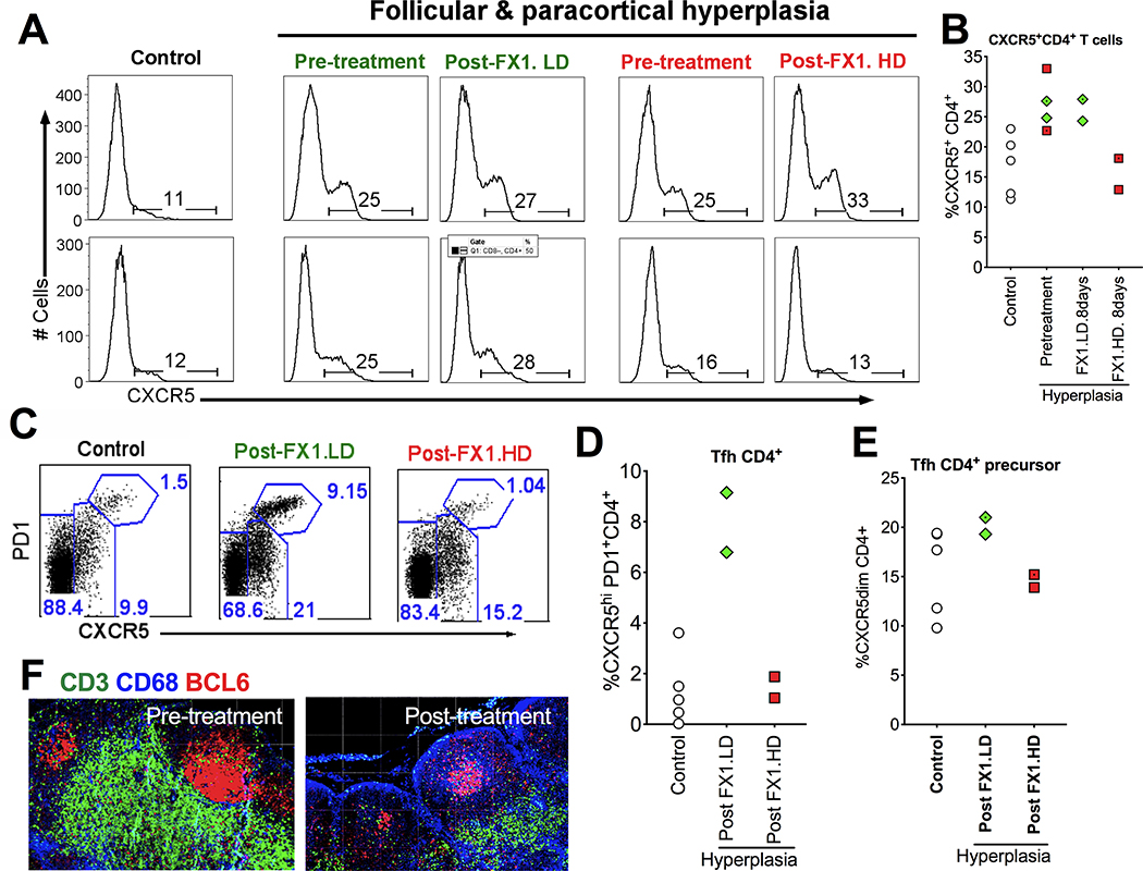
Figure 2. BCL6 BTB inhibitor (FX1) treatment reduced the frequency of lymphoid Tfh CD4+ T cells and its precursor cells.
(A-B) Representative histogram (A) and summary (B) showing the frequency of CXCR5+CD4+ T cells in the lymph node of healthy control macaques, macaques with lymphoid hyperplasia, and those macaques with lymphoid hyperplasia receiving an 8-day course FX1 treatment (except 7-day course FX1 treatment for one animal receiving FX1 at 25mg/kg). (C) Representative flow plot for the frequency of CXCR5hiPD1hi Tfh CD4+ T cells and its precursor cells (CXCR5dimCD4+) in the lymph node of four healthy control macaques, macaques with lymphoid hyperplasia, and those macaques with lymphoid hyperplasia receiving an 7- or 8-day course FX1 treatment at 25mg/kg. (D-E) The frequency of Tfh (D) and Tfh CD4+ precursor cells (E) in lymph nodes from four macaques with lymphoid hyperplasia at baseline and 48hr after an 7- or 8-day course FX1 treatment, as well as four healthy control macaques. (F) Representative images for the lymph node biopsies from the same adult macaques with lymphoid hyperplasia before (left) and 48hr (right) after FX1 treatment (25mg/kg). Anti-CD3 (green), anti-CD68 (blue), and anti-BCL6 (red) are presented. Images were captured at 200x magnification.

