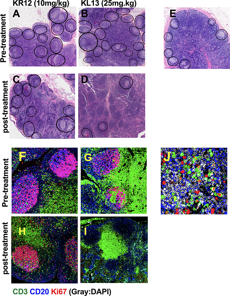
Figure 3. BCL6 BTB inhibitor (FX1) treatment reduced lymphoid hyperplasia and Ki67+ T cells in the germinal center.
Lymph node biopsies were collected from four adult macaques exhibiting lymphoid hyperplasia at baseline and 48hr after an 8-day course FX1 treatment. (A-D) H&E staining of lymph node biopsies at baseline (A&B) and after (C&D) 10mg/kg (C) and 25mg/kg (D) FX1 treatment for an 8-day course are presented. (E) Representative images for H&E stained lymph node tissue sections from healthy rhesus macaques. Images were captured at 100x magnification; Germinal center areas are highlighted in each section (see black ovals). (F-I) Immunofluorescence staining of the lymph node biopsies from macaques with lymphoid hyperplasia before treatment (F&G) and after receiving BCL6 FX1 treatment at 10mg/kg (H) and 25mg/kg (I). (J) Higher mangnification of baseline (pre-treatment) images are shown for the expression of Ki67 in the germinal center T cells. The tissue sections were stained with anti-CD3 (green), anti-CD20 (blue), anti-Ki67 (red) and DAPI (grey). Images were captured at 400x magnification.

