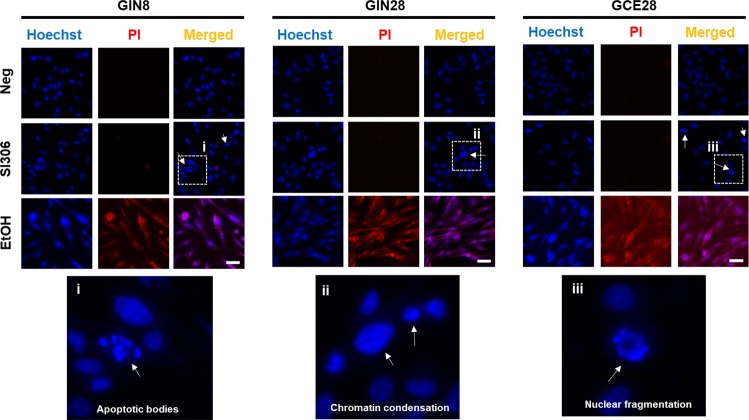
Figure 3.
Ho/PI staining of nuclei. Effect of SI306 kinase inhibitor on apoptotic features of cellular nuclei. Cells were dosed with 12.5 μM SI306, 70% ethanol (EtOH) or DMEM (Neg, negative control) for 48 h followed by nuclear staining with Hoechst 33342 and propidium iodide dyes. Scale bar indicates 30 μm. Images shown are representative of three sets of independent images. White arrows indicate the presence of apoptotic nuclei (chromatid condensation, apoptotic bodies).

