
FIGURE 4.
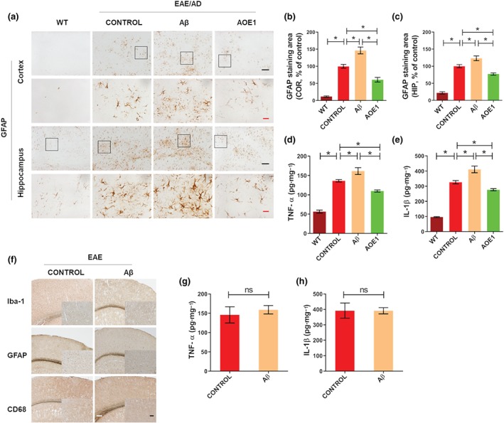
Immunization with Aβ42 increased astrocytosis and the levels of proinflammatory factors in the EAE/AD mice but did not exacerbate neuroinflammation in the EAE mice. (a) GFAP immunostaining for astrocytes in the brains of EAE/AD mice immunized with Aβ42 or AOE1. Scale bars: black, 100 μm; red, 20 μm. (b, c) Quantification of GFAP‐positive area in the cortex (b) and hippocampus (c). (d, e) The levels of TNF‐α (d) and IL‐1β (e) in the brain lysates of EAE/AD mice immunized with Aβ42 or AOE1 were detected by ELISA. (f) Iba‐1, GFAP, and CD68 immunostaining for gliosis in the brains of EAE mice immunized with Aβ42 or adjuvant. Scale bars: white, 200 μm; black, 100 μm. (g, h) The levels of TNF‐α (g) and IL‐1β (h) in the brain lysates of EAE mice immunized with Aβ42 or adjuvant were detected by ELISA. Data shown are means ± SEM, from n = 8 mice per group. *P < .05, significantly different as indicated; ns, not significantly different as indicated