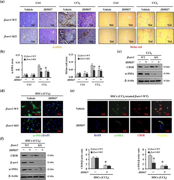
Figure 4.

β‐Arrestin1 was involved in the blockage of HSCs activation and liver fibrosis, by JD5037. (a) α‐SMA staining (brown) and Sirius red staining (red) were represented both in β‐arr1‐WT mice and in β‐arr1‐KO mice from CCl4‐ or olive oil‐treated (as Ctrl) mice, with or without JD5037 treatment (n = 6 per group). (b) α‐SMA area and Sirius red area from histological staining were determined. Data shown are means ± SD from n = 6 mice per group. *P < .05, significantly different from CCl4‐treated β‐arr1‐WT mice without JD5037. (c) Western blotting presented JD5037 ameliorated the levels of α‐SMA in CCl4‐treated β‐arr1‐WT mice (n = 6 per group). (d) JD5037 or β‐arr1 deficiency inhibited α‐SMA expression of hepatic stellate cells (HSCs) in CCl4‐treated mice. Nuclei (blue) were counterstained with DAPI (400×, n = 6 per group). (e) Co‐staining of α‐SMA (green) and CB1 receptors (CB1R; red) in primary HSCs dissociated from olive oil‐ and CCl4‐treated β‐arr1‐WT mice, respectively. Cell nuclei (blue) were counterstained by DAPI. (f) The indicated proteins from primary HSCs in the mouse models were detected by western blotting. The ratio of densitometry units of the normalized CB1 receptor/β‐actin and α‐SMA/β‐actin was also determined. Data shown are means ± SD from n = 6 mice per group. *P < .05, significantly different from primary HSCs from CCl4‐treated β‐arr1‐WT mice without JD5037
