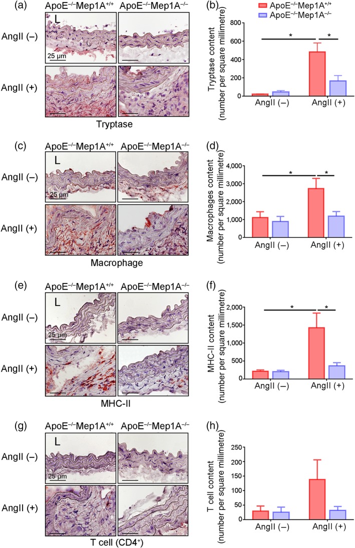
FIGURE 4.

Role of Mep1A in inflammation in abdominal aortic aneurysm (AAA). Representative images of the immunohistochemical analysis of (a) tryptase‐positive cells, (c) macrophages, (e) MHC‐II‐positive cells, and (g) CD4+ T cells in the mouse arterial wall. Immunohistochemical analysis of (b) tryptase‐positive cells, (d) macrophages, (f) MHC‐II‐positive cells and (h) CD4+ T cells in the arterial walls of the experimental mice of the indicated groups. The bar graphs present the positive cells per square millimetre. Data represent the mean ± SEM. Groups: ApoE−/−Mep1A+/+ mice treated with PBS (n = 5), ApoE−/−Mep1A−/− mice treated with PBS (n = 6), ApoE−/−Mep1A+/+ mice treated with Ang II (n = 5) and ApoE−/−Mep1A−/− mice treated with Ang II (n = 6). Two‐way ANOVA was conducted to examine the differences among groups in all data from experimental AAAs. *P <.05 was considered statistically significant. L, lumen
