Figure 1.

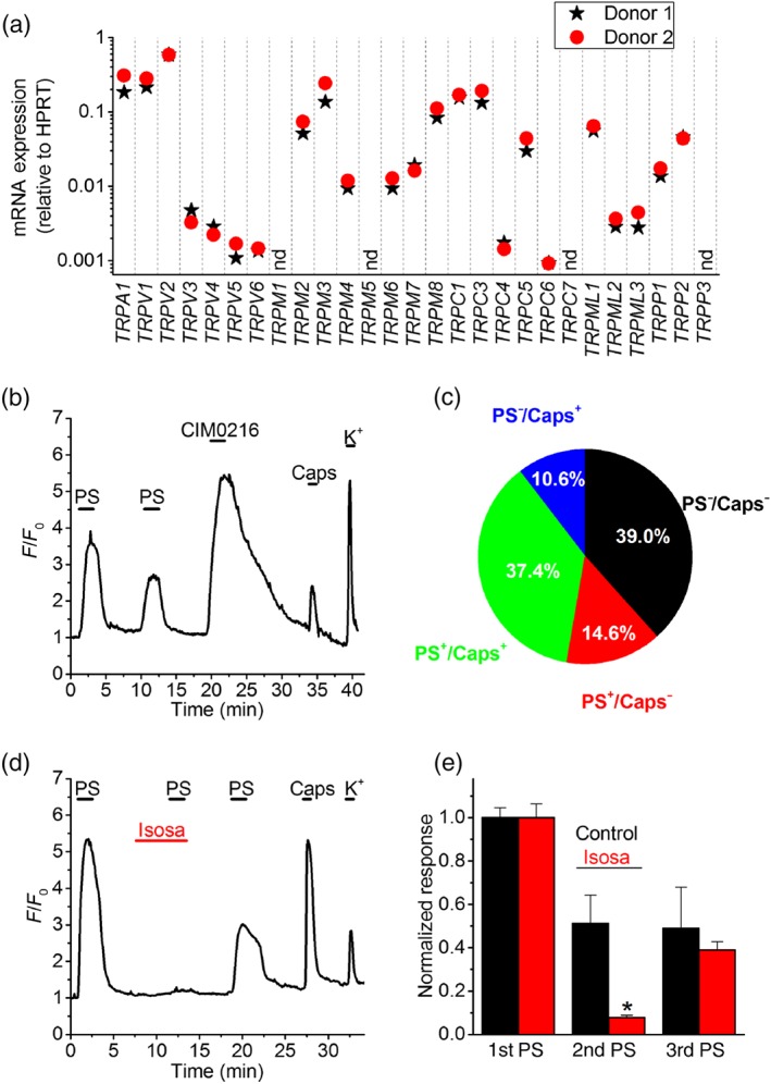
Functional TRPM3 expression in human dorsal root ganglion (DRG) neurons. (a) Expression levels of TRP channel mRNA relative to HPRT in DRG neurons from two donors, determined using RT‐qPCR. Access to fresh human DRG tissue is scarce, hence the limited number of samples in this exploratory experiment. nd, not detected. (b) Representative example of changes in Fluo‐8‐fluorescence in human DRG neurons in response to the TRPM3 agonists pregnenolone sulphate (PS; 50 μM) and CIM0216 (10 μM) and to the TRPV1 agonist capsaicin (Caps; 200 nM). A high K+ solution was applied at the end of the experiment to confirm neuronal excitability. A trace showing the mean ± SEM of the changes in Fluo‐8‐fluorescence for all cells in this particular experiment is provided in Figure S1A. (c) Pie chart showing the distribution of neurons responding to PS, capsaicin or both (n = 206 hDRG neurons). (d) Representative example showing the reversible inhibition of PS‐induced responses in hDRG neurons by the selective TRPM3 antagonist isosakuranetin (Isosa; 20 μM). A trace showing the mean ± SEM of the changes in Fluo‐8‐fluorescence for all cells in this particular experiment is provided in Figure S1B. (e) Normalized responses to repeated PS applications. Neurons were stimulated three times with PS, and the second application occurred in the presence of either isosakuranetin (10 μM; n = 50) or vehicle control (n = 39). *P < .05 (two‐way repeated measures ANOVA with Tukey's post hoc test) [Colour figure can be viewed at http://wileyonlinelibrary.com]
