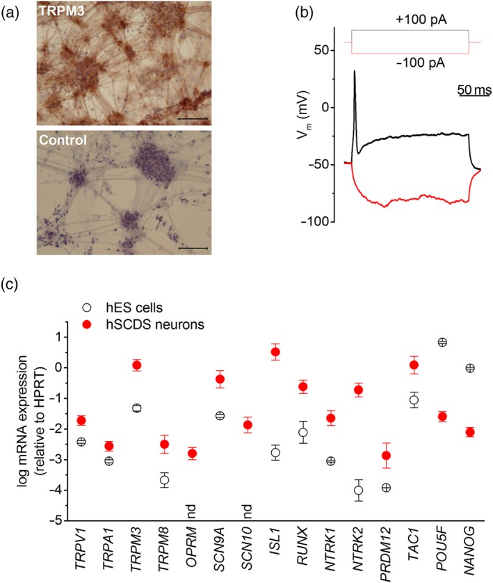
Figure 2.

Properties of human stem cell‐derived sensory neurons (hSCDS) neurons. (a) Brightfield image of immunohistochemically stained hSCDS neurons with anti‐TRPM3 antibody and negative control (only secondary antibody) showing a neuronal morphology with cell body clusters interconnected by neurites. Brown peroxidase‐based TRPM3 staining is visible in cell bodies as well as neurites. Scale bar: 500 μm. (b) Whole‐cell current‐clamp recordings on hSCDS neurons demonstrate the ability to fire action potentials upon current injection. (c) RT‐qPCR comparing relative expression of sensory TRP channels, nociceptor marker genes, and pluripotency genes between human embryonic stem (hES) cells (black) and hSCDS neurons (red). Results represent mean ± SEM from five independent experiments with three technical replicates each. nd, not detected in hES cells human embryonic stem cells [Colour figure can be viewed at http://wileyonlinelibrary.com]
