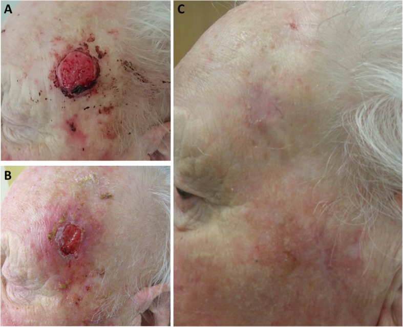
Fig. 1.

Clinical case of a 94-year old male patients who received radiotherapy for a well-differentiated squamous cell carcinoma. a Pre-therapeutic situation in November 2015 showing an ulcerating and bleeding skin lesion on the left temple. Radiotherapy was performed using electron beams with a total of 51 Gy (27 Gy applied in 9 fractions followed by 24 Gy in 6 fractions). b Irradiation-induced dermatitis (grade 2) in December 2015 after completion of treatment. c Follow-up consultation in April 2016 showing a complete clinical response with no high-grade radiotherapy-related toxicities
