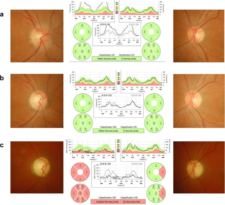
Fig. 1.
Fundus photography and optical coherence tomography results at each episode. a, At the first episode of optic neuritis in the right eye (acute optic neuritis, OD), there was optic disc swelling in the right eye with no abnormality in the left eye. b, Seven months after the right optic neuritis, the patient developed acute painless visual loss in the right eye (LHON, OD). There was no optic disc swelling in the right eye with mild temporal disc pallor. c, 22 months after the right optic neuritis and 4 months after the new development of left optic neuropathy (4 months after bilateral development of LHON), there was global disc pallor in the right eye and temporal disc pallor in the left eye

