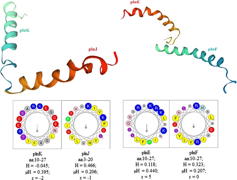
Fig. 4.
3D structures of PlnJK and PlnEF plantaricin peptides of SF9C strain predicted by the homology modelling and their helical wheel projections analysed by HeliQuest. aa—the amino acid residues (the one-letter code for amino acids is used); yellow—hydrophobic residues; purple—serine and threonine residues; dark blue—basic residues; red—acidic residues, pink—asparagine and glutamine residues; grey—alanine and glycine residues, light blue—histidine residues; green—proline residues; H—hydrophobicity; μH—hydrophobic moment; z—net charge (calculated at pH = 7.4, under the assumption that histidine is neutral and that the N-terminal amino group and the C-terminal carboxyl group of the sequence are uncharged)

