. 2020 Mar 25;11(5):832–838. doi: 10.1021/acsmedchemlett.9b00639

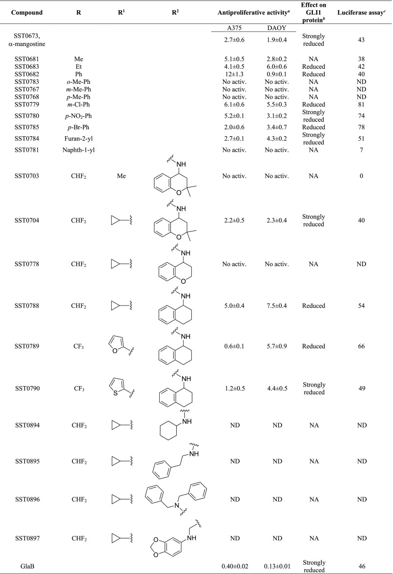
Table 1. Structure and Antiproliferative Activity of Compounds That Affect GLI1.

graphic file with name ml9b00639_0010.jpg

graphic file with name ml9b00639_0011.jpg

a

Expressed as IC50 values (micromolar concentrations) calculated using GraphPad prism, version 6, from triplicate experiments in melanoma (A375) and medulloblastoma (DAOY) cells. ND: not determined. No activ.: no effects on proliferative activity.

b

GLI1 protein level was determined by Western blotting (see Figure 3 for details) in murine NIH3T3 cells treated with SAG (100 nM) and each compound (5 μM) for 48 h. NA: not affected.

c

Luciferase assay in murine NIH3T3 cells. Data are represented as percentage of inhibition. Cells treated with SAG were equated to 100% (see Figure 4 for details). ND: not determined.