Figure 1.
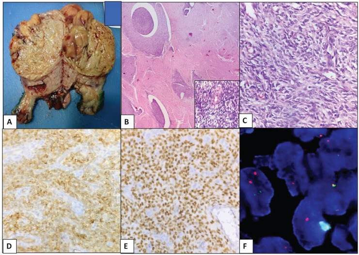
Low-grade endometrial stromal sarcoma. (A) Gross photo: well-circumscribed, yellow to tan fleshy cut surface. (B) Islands of tumour cells with tongue-like growth pattern without stromal response (H&E, ×100). Inset – delicate spiralling network of arterioles (haematoxylin and eosin [H&E], ×400). (C) Uniform small cells with minimal atypia (H&E, ×400). (D) Diffuse strong positive for CD10 (×400). (E) Diffuse positive for ER (×400). (F) JAZF1-SUZ12 dual fusion probe detecting t(7;17)(p15;q11.2) rearrangement. CD10 indicates cluster differentiation 10; ER, oestrogen receptor.

