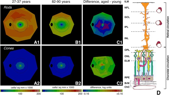
Fig. 1.
Aging of the human photoreceptor mosaic and the outer retinal neurovascular unit. A-C Rod vulnerability and cone resilience in healthy aging [34, 35]. Topography of rods and cones determined from computer-assisted cell counts in flat-mounts of human retina [3]. Maps are shown as a fundus of a left eye. Black oval, optic nerve; black ring, outer limit of Early Treatment of Diabetic Retinopathy Study grading grid. A1, A2. Rods and cones in 27–37-year-old donors. B1, B2. Rods and cones in 82–90-year-old donors. C1, C2. Log mean difference in cell density between younger and older adults. C1. Difference in rod density between younger and older adults is greatest at 0.5 mm to 3 mm from fovea (parafovea and perifovea). Purple signifies that aged eyes had 31% fewer cells than young eyes. C2. Log mean difference in cone density between younger and older adults is small and inconsistent, indicated by the yellow-green map. D Outer retinal neurovascular unit and retinoid re-supply to cone and rod photoreceptors. Shown are rods (R), cones (C), Müller glia (M), RPE, and vascular endothelium of the choriocapillaris (ChC) and capillary plexuses of the retinal circulation. We hypothesize that to the photoreceptors, AMD is a disease of the retinoid re-supply route. Vitamin A delivered from plasma is rate-limiting for recovery of sensitivity by rods. Rods need choriocapillaris, Bruch’s membrane, and RPE, whereas the cones have these, plus an additional second delivery route, via Müller glia and the retinal circulation. RMDA assesses how pathology in the choriocapillaris, Bruch’s membrane, and RPE complex impacts rods. It is expected that cone-mediated vision will be resilient. Retinal layers: ILM, inner limiting membrane; NFL, nerve fiber layer; GCL, ganglion cell layer; IPL, inner plexiform layer; INL, inner nuclear layer; OPL, outer plexiform layer; ONL, outer nuclear layer; ELM, external limiting membrane; RPE, retinal pigment epithelium; BrM, Bruch’s membrane; ChC, choriocapillaris

