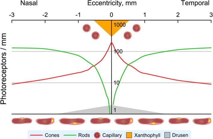
Fig. 2.
Visual neuroscience of human retinal aging and age-related macular degeneration.Schematic shows log cone and rod density [30], choriocapillaris and retinal vessels, and helpful/ harmful factors for photoreceptor function and survival in the human macula. Soft drusen and basal linear deposits (gray) accumulate between choriocapillaris and retinal cells. They are thickest and confer highest risk for progression in central macula. Xanthophyll carotenoid pigment (orange) is highest in the foveal center (shown) with lateral extensions into the plexiform layers (not shown), plausibly attributable to the distribution of protective Müller glia [34]. The retinal capillaries and choriocapillaris are shown

