Figure 1.
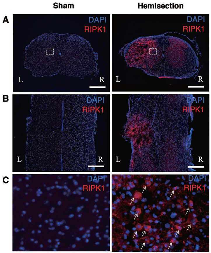
Upregulation of RIPK1 expression in the spinal cords after hemisection. Immunostaining of RIPK1 reveals that the uninjured cords showed no obvious expression of RIPK1 in the transverse (A) and horizontal (B) sections. At 3 days after the hemisection, the RIPK1 expression on the injured side (L) increased compared with that on the contralateral side (R) in the transverse (A) and horizontal (B) sections. Scale bars: 500 μm. High-magnification views (C) of boxed areas in (A). RIPK1-positive cells (arrows) increased at the lesion site (C). RIPK1 indicates receptor-interacting protein kinase 1.

