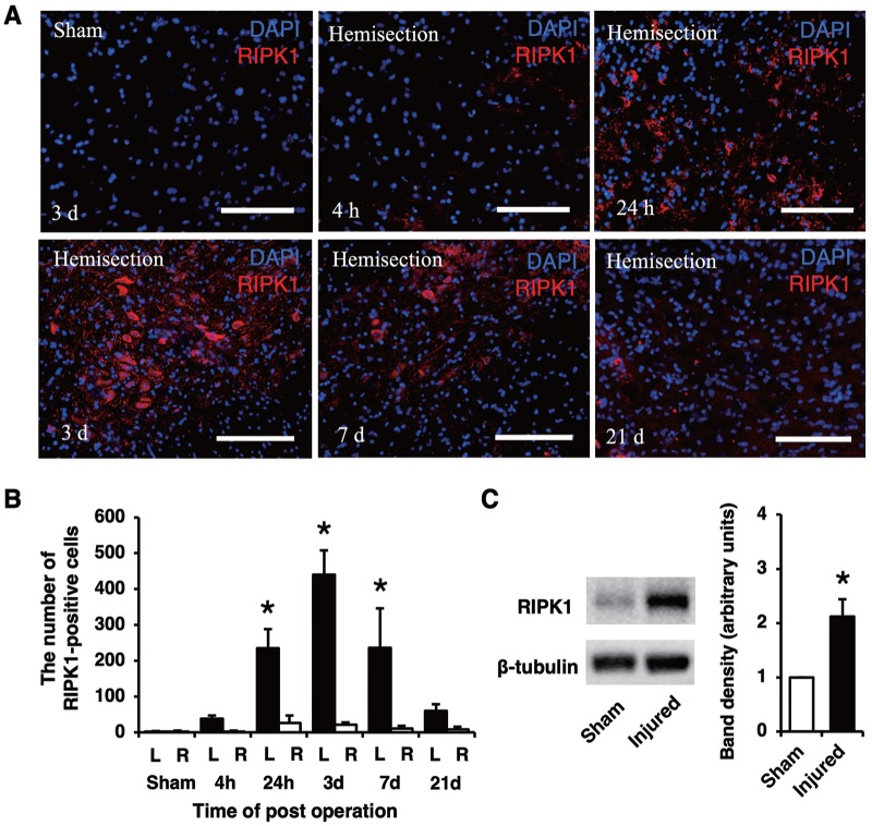
Figure 2.
Immunostaining of RIPK1 on the injured side at different time points. The population of RIPK1-expressing cells at 24 hours and 3 and 7 days were larger than those at the other time points (A). Scale bars: 100 μm (A). The number of RIPK1-positive cells was significantly higher on the injured side (L) than that on the contralateral side (R) and in the sham group at 24 hours and 3 and 7 days (B). Western blotting showed that the RIPK1 protein expression increased in the injured spinal cord (C). A quantitative analysis of the band density revealed that the level of RIPK1 proteins in the injured spinal cord was significantly higher than that in the uninjured spinal cord samples. All values are presented as mean ± SD. *P < .05, n = 3 and 5 per each group in (B) and (C), respectively. RIPK1 indicates receptor-interacting protein kinase 1.

