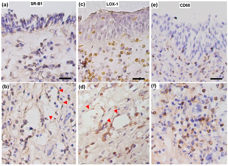
Figure 4.
Representative immunohistological images showing SR-B1 (a,b), LOX-1 (c,d), and CD68 (e,f) expression in ethmoid sinus mucosa sampled from a CRSwNP patient. Vascular endothelial cells (arrowheads) are stained positively both for SR-B1 and LOX-1. In contrast, numerous submucosal inflammatory cells show intense positive staining for LOX-1 compared to that for SR-B1. Scale bar: 20 μm.

