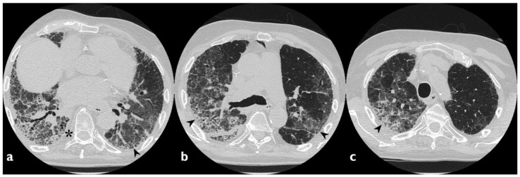
Figure 4.
A case of Amiodarone-Induced Lung Toxicity (AILT). Axial scan passing through the bases (a), through the origin of the pulmonary artery (b), and through the apices (c). Reticulations, traction bronchiectasis, and widespread areas of GGO are shown in panels a–c (black arrowheads); parenchymal alterations have central and peripheral distribution. At the bases, in the subpleural field, an initial honeycomb pattern is appreciable (asterisk).

