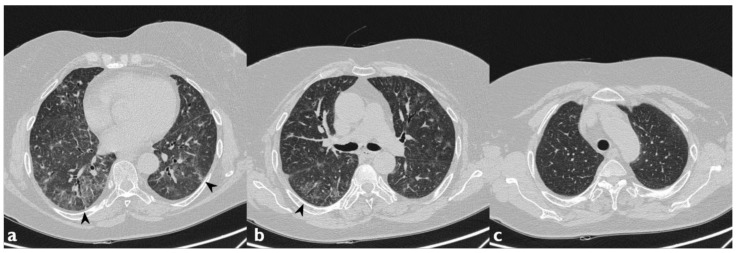
Figure 8.
Another case of AILT. Axial scan passing through the bases (a), through the origin of the pulmonary artery (b), and through the apices (c). Interstitial disease with NSIP pattern. Diffuse increase in density of the lung parenchyma with a GGO appearance (black arrowheads indicate the previous findings).

