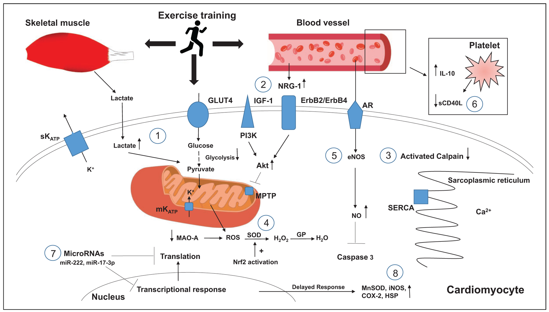
Figure 1. Proposed mechanisms of exercise cardioprotection.

1. Lactate from skeletal muscle and intracellular cardiomyocytes provide substrate for ATP generation during anaerobic glycolysis.
2. NRG-1 levels increase binding to erbB2/erbB4, activating the PI3K/Akt pathway that inhibits MPTP opening during cellular hypoxia.
3. Intracellular calcium load is decreased, leading to decreased activation of calpain.
4. Decreased levels of MAO-A and increased SOD activity through activation of Nrf2 activation.
5. Endothelial eNOS and intracellular NO levels rise, inhibiting caspase 3 activity.
6. Decreased circulating sCD40L and increased production of IL-10.
7. Increased levels of miRNAs such as miR-222 and MiR-17–3p lead to inhibition of transcriptional and translation responses deleterious to cardiomyocyte function.
8. Genetic modification leads to de novo synthesis of effector proteins during second window of protection.
