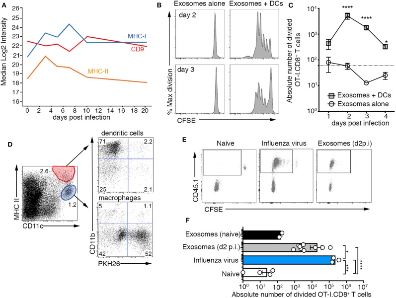
Figure 3.
Airway exosomes contain influenza virus antigen and are capable of initiating a cellular immune response. (A) Proteomic analysis measuring levels of CD9, MHC II, CD74, and MHC I expressed by exosomes isolated from BALf over the course of an influenza virus infection. (B,C) Exosomes purified from the BALf of mice infected 1–4 days prior with 104 PFU of X31-OVA (1) were cultured with 5 × 10⁴ CFSE-labeled OT-I CD8+ T cells with or without 1 × 10⁴ DCs. T cell proliferation was measured 60 h later. (B) Representative OT-I.CD8+ T-cell proliferation profiles with exosomes recovered from the BALf on days 2 and 3 p.i., cultured with or without DCs. (C) The absolute number of divided OT-I.CD8+ T cells. Data pooled from 3 to 9 independent experiments. Symbols represent the mean ± sem (two-way ANOVA, Sidak's multiple comparison). Dotted line represents the background level of T-cell division. (D) PKH26-labeled exosomes were intranasally administered into mice and 1 h later the lungs were recovered and the proportion of DCs and macrophages that had engulfed PKH67+ exosomes was profiled. (E) Mice injected with 2 × 10⁶ CFSE-labeled OT-I.CD8+ T cells were intranasally administered either saline (naïve) or x31-OVA (1) or 0.75 mg of exosomes recovered from the BAL of naïve mice or mice infected with x31-OVA (1) 2 days earlier and the absolute number of divided OT-I cells in the mLN was measured 4 days later. (E) Representative flow cytometry profiles gated on CD8+ T cells. (F) Graph depicts the absolute number of divided cells. Data pooled from 4 independent experiments (one-way ANOVA, Dunnett's multiple comparison). *p < 0.05, ***p < 0.001, ****p < 0.0001.

