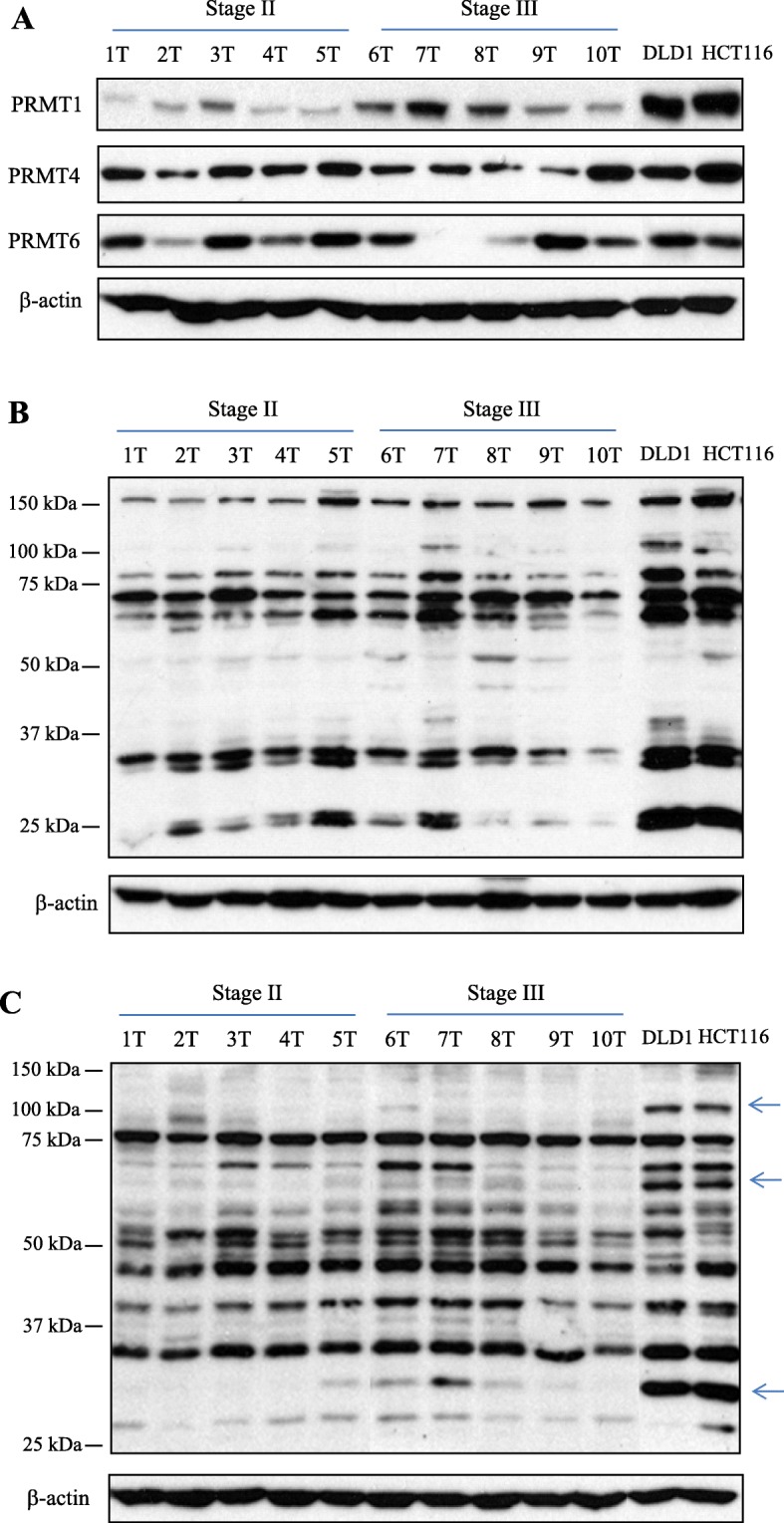
Fig. 1.

Expression of type I PRMTs, MMA- and ADMA-containing proteins in CRC tissues from patients as well as established CRC cell lines. a Equal amount of each CRC tissue extract from 10 patients and DLD1 and HCT116 cell lysates were subjected to Western blot analysis with the respective type I PRMT antibodies. b Arginine monomethylation status was examined with MMA-specific antibody. The product was conjugated to protein A agarose beads in PTMScan MMA motif kit. c Asymmetric arginine dimethylation status was examined with ADMA-specific antibody. The product was also used for IAP in a PTMScan ADMA motif kit. Arrows indicates highly expressed ADMA-containing proteins in two CRC cell lines, compared to those of 10 CRC tissues
