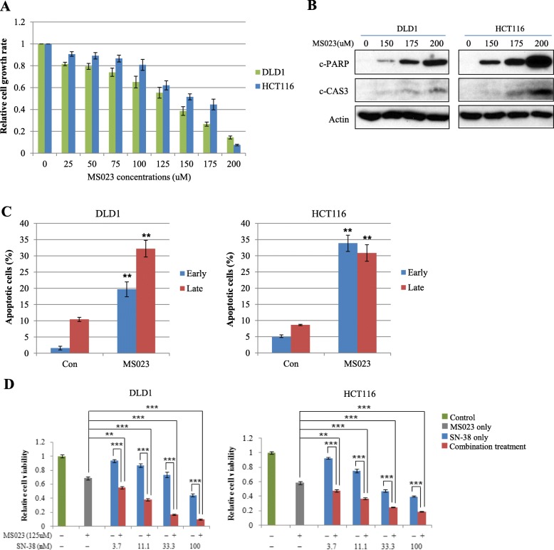
Fig. 4.
Treatment of type I PRMT inhibitor on CRC cells significantly increases CRC cell apoptosis and suppress CRC cell proliferation by SN-38. a DLD1 and HCT116 cells were seeded at a density of 5000 cells/well in 96 well plates. After 24 h, cells were treated with increasing concentrations of MS023 as indicated and cell viability was determined by WST-1 based assay. b To examine the induction of CRC cell apoptosis by MS023, two CRC cell lines were seeded at a density of 3 × 10⁵ cells/well in 6well plates. After 24 h, cells were treated with increasing concentrations of MS023 as indicated for 48 h. Induction of cleaved forms of caspase 3 and PARP proteins was analyzed by Western blot. c With the same condition shown in panel (b), mean annexin-V and propidium iodide fluorescence intensities in early and late apoptotic cell populations were quantified in three independent experiments. Representative scatter plots of each cell line are presented in Additional file 8: Figure S4. **, p < 0.01. d DLD1 and HCT116 cells were exposed to increasing concentrations of SN-38 with a fixed MS023 concentration as indicated above for 48 h. Cell viability was measured by WST-1 assay. Error bars represent the mean ± SD of three independent experiments. The Student’s t-test was performed between cells exposed to MS023 or SN-38 alone and cells exposed to MS023 plus each concentration of SN-38 in two CRC cell lines. **, p < 0.01; ***, p < 0.001

