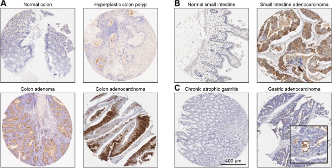
Fig 5. Immunohistochemical detection of NOX1 protein in tumors and corresponding non-neoplastic tissue of the same origin.
Representative tumors/lesions and normal/non-neoplastic tissue pairs from a tissue microarray are shown. (A) Normal and abnormal colon, (B) normal and malignant small intestine, and (C) chronic atrophic gastritis and malignant gastric tissue. All images were taken at 10X digital magnification. The scale bar represents 400 μm.

