
Figure 5.
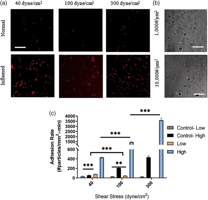
Adhesion of aICAM‐1 functionalized nanoparticles (NPs) to activated Endothelial cells (ECs) under high shear conditions. (a) Representative fluorescent confocal microscopy images following perfusion experiments over ECs. The images show higher adhesion of aICAM‐1 (10,000 copies/μm2) NPs to activated cells at the examined wall shear stresses: (i.e., 40, 100, and 300 dyne/cm2). Scale bar: 50 μm. (b) Confocal microscopy images comparing the adhesion of aICAM‐1 functionalized NPs at two surface densities (1,000 & 10,000 #/μm2) to activated ECs phase image) at a WSS of 300 dyne/cm2. High‐density aICAM‐1 particles show greater adhesion to cells. Scale bar: 50 μm. (c) Quantification of the adhesion rate of aICAM‐1‐NPs coated with two densities to activated ECs under flow at different levels of wall shear stress (i.e., 40,100, and 300 dyne/cm2). The EC were stimulated by TNF‐α for 6 hr