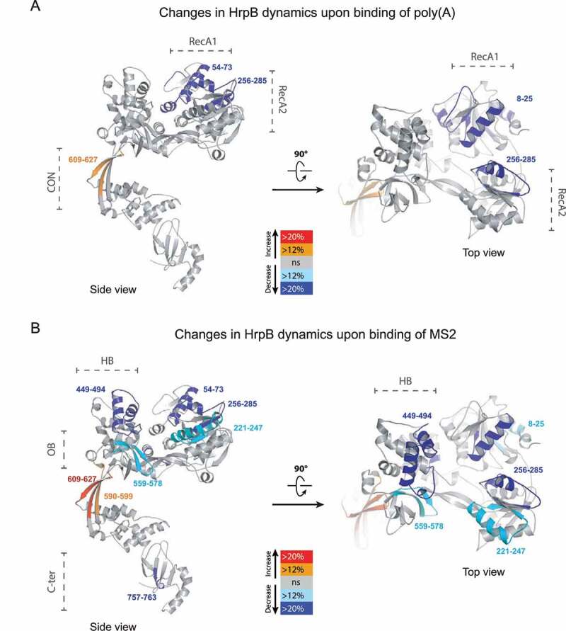
Figure 8.

Mapping of RNA responsive region on HrpB structure. Peptides with significant changes in presence of poly(A) (A) or MS2 (B) are coloured on the ribbon diagram of the P. aeruginosa HrpB structure (Supplementary Figure S9) using PyMOL and according to the colour scheme shown (red and orange for increased hydrogen-deuterium exchange [HDX], cyan and blue for decreased HDX).
