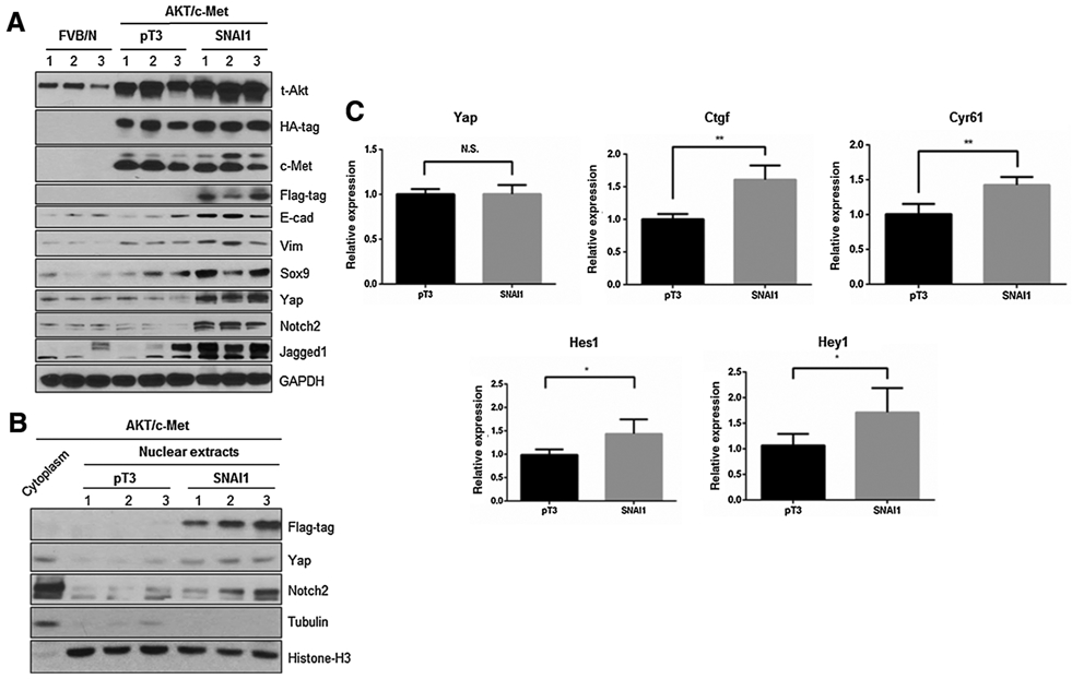
Figure 4.

Yap signaling activity is increased in AKT/c-Met/SNAI1 mouse liver tissues. A, Western blot analysis of relative protein expression in AKT/c-Met/pT3 and AKT/c-Met/SNAI1 mouse liver tissues. B, Western blot analysis was performed in nuclear and cytoplasmic protein extracts from FVB/N normal mouse livers and AKT/c-Met/pT3, AKT/c-Met/SNAI1 mouse liver tumor samples. C, Detection of Yap and Notch target genes by qRT-PCR in AKT/c-Met/pT3 and AKT/c-Met/SNAI1 mouse liver tissues. *, P < 0.05; **, P < 0.01. N.S., not significant.
