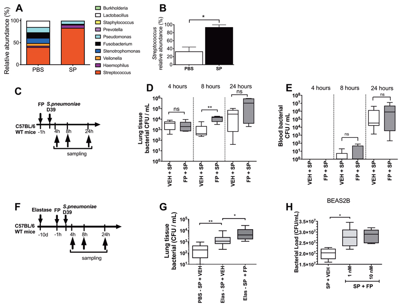
Figure 2. Fluticasone propionate impairs pulmonary clearance of Streptococcus pneumoniae.
(A-B) Lung microbiota was evaluated by 16S rRNA sequencing in lung tissue from mice challenged with S. pneumoniae (SP) and PBS treated controls at 8 hours post-challenge. (A) Relative abundance of the top ten operational taxonomic units (OTUs) (B) Relative abundance of Streptococcus. (C) Experimental outline. C57BL/6 mice were treated with 20ug FP or vehicle DMSO control and additionally infected with S. pneumoniae D39. Bacterial loads in (D) lung tissue and (E) blood were measured at the indicated timepoints post-infection by quantitative culture. (F) Experimental outline. C57BL/6 mice were treated intranasally with a single dose of elastase or PBS as control. Ten days later, mice were treated intranasally with fluticasone propionate (20μg) or vehicle DMSO control and challenged with S. pneumoniae D39. (G) Bacterial loads were measured at 8 hours post-infection. (H) BEAS-2B cells were treated with 1 or 10nM fluticasone propionate, stimulated with S. pneumoniae D39 and bacterial loads were measured in cell supernatants by quantitative culture at 24 hours post-infection. Bacterial load data are displayed as box and whisker plots showing median (line within box), IQR (box) and minimum to maximum (whiskers). Experiments comprise n=6-8 mice/group, representative of at least two independent experiments. Data analyzed using Mann Whitney U test or one-way ANOVA with Bonferroni post-test. n.s. non-significant *p<0.05 **p<0.01 ***p<0.001.

