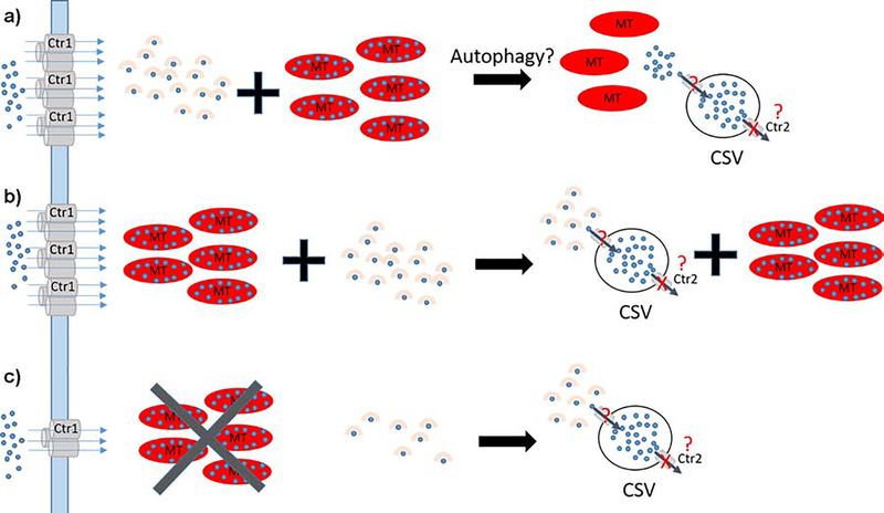
Figure 3. Possible models of CSV formation.
a) Model 1: Imported Cu (blue) is bound by chaperones or high affinity peptides (beige) delivered to target proteins or stored by MTs (red ovals). CSVs may be formed as a consequence of autophagy that initiates Cu release from MT and subsequent import into CSVs resembling what was described for MT3 [13]. Export is blocked because no Ctr2 is present. b) Model 2: In contrast to a) CSVs are populated by Cu from Cu chaperones or Cu-bound peptides. c) CSV formation in MT knockout brains. Model 2 can explain our XFM observations: MT knockouts reduce the cellular Cu storage capacity and signal for reduced Ctr1 expression at the plasma membrane. CSVs are populated with Cu as described in b).

