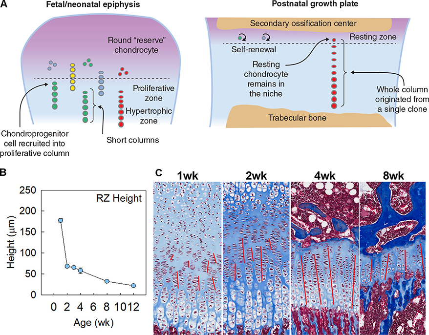
Figure 3. Establishment of resting zone stem cell niche.
In fetal and neonatal life, some chondroprogenitor cells in the epiphysis are recruited into the proliferative columns, leading to their gradual depletion early on. However, after the formation of secondary ossification center, the balance appears to shift toward resting chondrocyte self-renewal, leading to formation of long columns from single clones (A). This is consistent with observations that resting zone height in tibia decreases drastically after 1 week in mice (B), and a tendency to find clusters of short columns in growth plate histology of 1- and 2-wk old mice, which gradually transition to more continuous straight columns in older animals, as indicated by the red lines (C).

