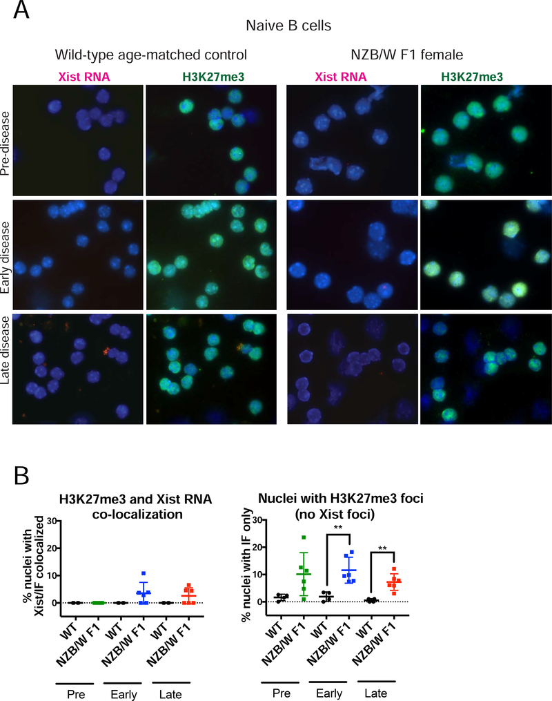
FIGURE 3. NZB/W F1 naïve B cells have more H3K27me3 foci compared to wildtype (C57BL/6; BALB/c) cells.
(A) Sequential Xist RNA FISH (left, red) followed by immunofluorescence (right, green) for H3K27me3 for naïve CD23+ B cells from wildtype age-matched mice (n = 4) and NZB/W F1 B cells (n = 6) for each disease category (Supplementary Figure 1). Arrowheads denote H3K27me3 foci; DAPI nuclear counterstain is shown in blue. Representative cells collected from multiple independent experiments are shown. (B) Quantification of co-localization of Xist RNA signals and H3K27me3 foci (left) and nuclei containing just H3K27me3 foci (right). (Left) The percentage of cells with detectable Xist RNA clusters overlapping with a focus of H3K27me3 was determined for each animal. (Right) The percentage of cells containing just a focus of H3K27me3 without any detectable clustering of Xist RNA is shown. The mean across replicates and standard deviation of the mean is shown. Significance was determined by unpaired t test. **p<0.01. Total nuclei counted: Pre-disease = 539, Pre-WT = 342, Early-disease = 432, Early-WT = 358, Late-disease = 511, Late-WT = 342.

