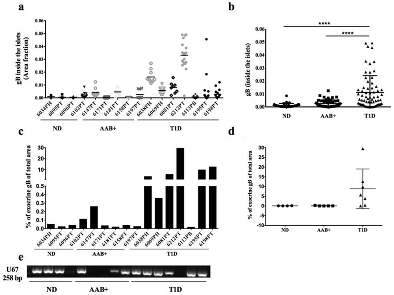
Figure 2. gB is more frequently detected in the pancreas of T1D donors.
Quantification of gB expression inside the islets (a, b) or in the exocrine (c, d) from non-diabetic donors (n=4), AAB+ donors (n=5) and donors with T1D (n=7). Each dot represents an islet and is presented as the positive fraction of the total islet area (a, b) or percentage of gB positive of total area (c). For each case, up to 10 islets were quantified. In (d), each dot represents a donor. (e) DNA was extracted from ND, AAB+ and donors with T1D, and U67 viral gene (258bp) was detected by nested PCR. ND: Non diabetic; AAB+: Auto-antibody positive; T1D: Type 1 diabetes.

