Figure 2.
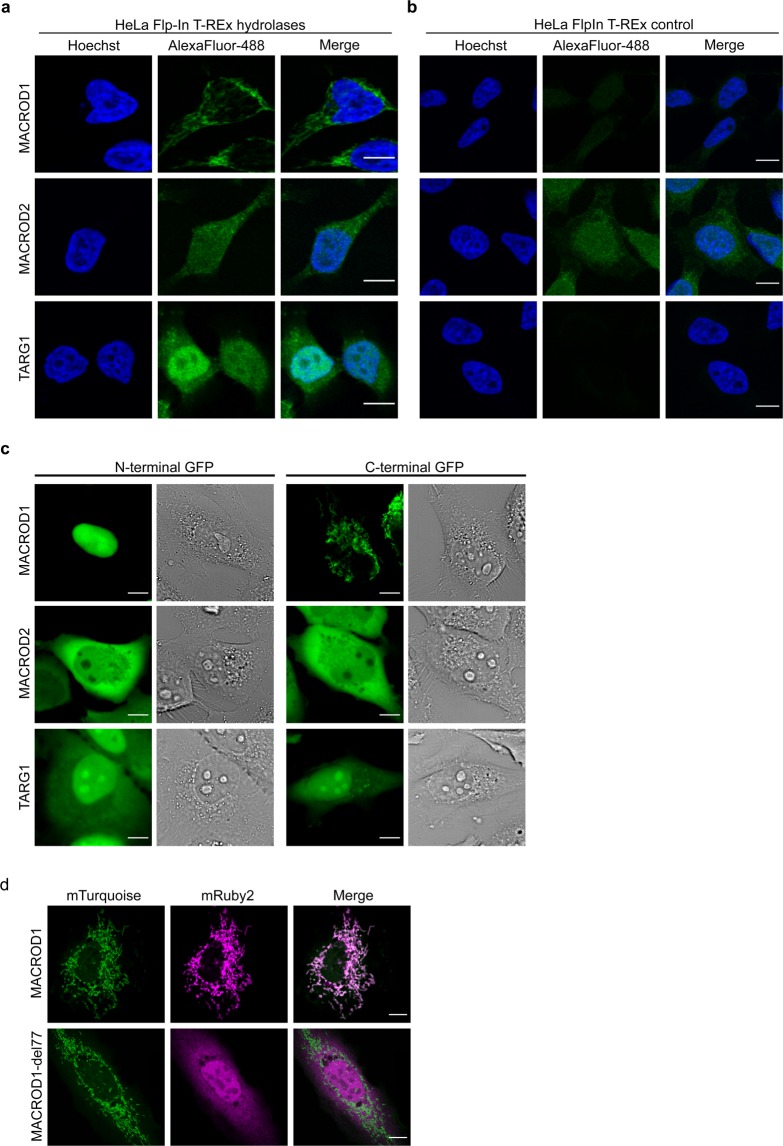
MACROD1, MACROD2 and TARG1 are differentially localised. (a) HeLa Flp-In T-REx cells overexpressing MACROD1, MACROD2 or TARG1 were treated overnight with 100 ng doxycycline/ml, fixed with PFA, stained with primary antibodies (monoclonal antibodies used: MACROD1 (28C11), MACROD2 (18D12), TARG1 (3A5)), visualised using an AlexaFluor488-coupled secondary antibody and analysed with confocal microscopy. (b) HeLa Flp-In T-REx control cells were treated and analysed as in panel (a). (c) HeLa cells were transfected with the indicated N- or C-terminally GFP-tagged constructs and analysed using live-cell confocal microscopy. (d) HeLa cells were transfected with constructs expressing mTurquoise2 targeted to mitochondria and either mRuby2-labeled MACROD1 full-length construct or an N-terminal truncation lacking amino acids 1–77, followed by live-cell confocal imaging. Scale bars represent 10 µM.

