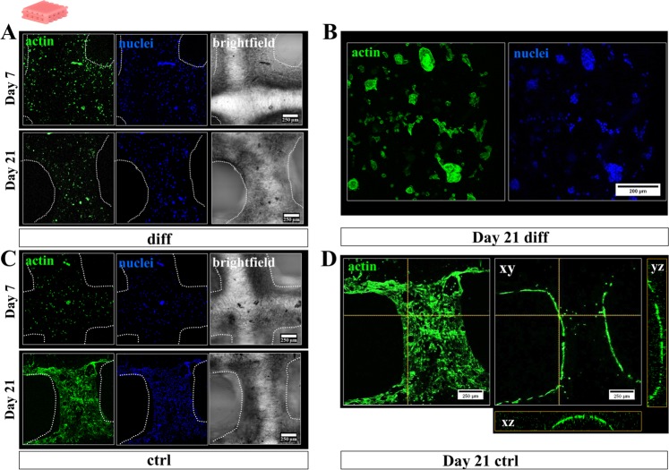
Figure 5.
Cytoskeletal morphology and distribution of hCh inside 3D plotted monophasic algMC scaffolds. Fluorescence staining of cell nuclei (blue: DAPI) and cytoskeletal F actin filaments (green: phalloidin) in diff and ctrl condition. (A,B) Cells provided with their respective chondrogenic factors remained inside the 3D matrix of the algMC strands after 7 and 21 days, starting to form chondrogenic clusters (B) after 3 weeks. (C,D) Cells in ctrl cultivation condition started to migrate out of the strand core towards the gel surface colonizing the entire strand. (D) Orthogonal views from additionally projected yz and xz perspective on the cell distribution in orthogonal layers of the strand.

