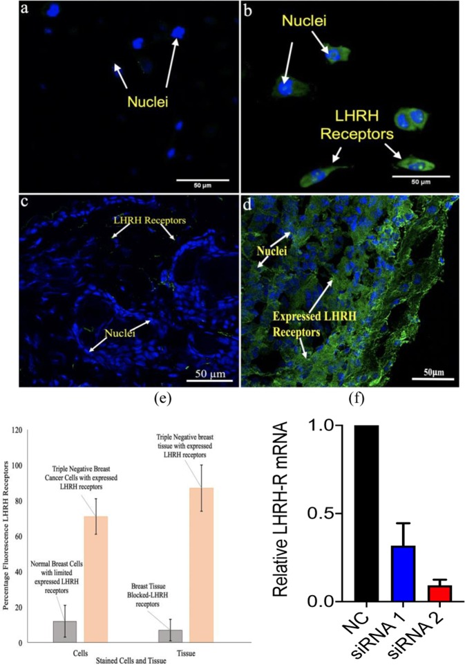
Figure 3.
Confocal fluorescence images showing the expression of LHRH receptors (green stains) of (a) non-tumorigenic epithelial breast cell line (MCF 10 A) (b) Triple negative breast cancer cells (MDA-MB 231) (c) Blocked LHRH antibody receptors on triple negative breast tissue (d) Stained LHRH triple negative breast tissue at 40 x magnification (e) Quantified fluorescence LHRH receptors in cells and tissue of TNBC. (f) Detection of LHRH-R knockdown by RT-qPCR.

