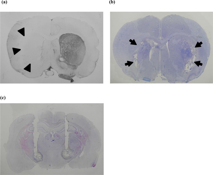
Figure 1.
Histological verification of animal models of hyperkinetic movement. (a) Tyrosine hydroxylase immunohistochemistry showing unilateral deprivation of dopaminergic innervation of the striatum (arrowheads) in a parkinsonian rat with levodopa-induced dyskinesia. (b) Bilateral striatal lesions (arrows) in a rat with hyperkinesia after direct intrastriatal 3-NP injection shown by cresyl violet staining. (c) Cresyl violet staining showing trajectories of the combined microinjection cannula/stimulation electrode in the bilateral subthalamic region in a levodopa-induced dyskinesia parkinsonian rat.

