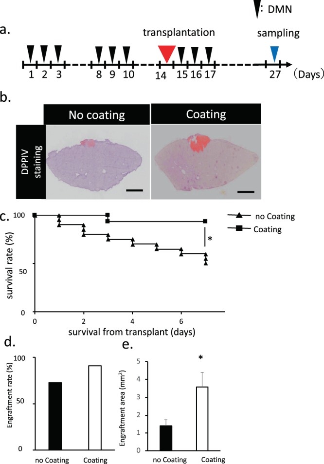
Figure 2.

Transplantation methods of fetal liver tissue onto the liver surface. (a) Schematic of the transplantation of a fetal liver tissue coated with a coating agent. After 27 days of first DMN injection, the samples were collected and analyzed. (b) Histological analysis of liver tissue with a coating agent covering the liver surface before transplantation. DPPIV-stained living tissue is colored in red. Scale bar: 2 mm. (c) Measurement of the survival rate of fetal liver tissue transplantation after coating agent utilization. The survival rate of the transplanted group without coating agent was also monitored as a control. Data are mean ± SE. *P < 0.01, log rank test; no coating: n = 20, coating: n = 15. (d,e) Measurement of the engraftment rate and area of fetal liver tissue transplantation after coating agent utilization. The engraftment rate and area in the transplanted group without coating agent was also monitored as a control. Data are mean ± SE. *P < 0.05, vs. wild-type Mann-Whitney U test, n = 11.
