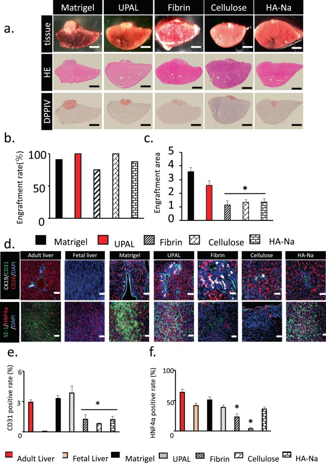
Figure 3.
Histological assessment of the transplanted fetal liver tissue in five coating agent groups. (a) Histological analysis of the fetal liver tissue that was engrafted on the surface of the middle lobe of the liver and covered by a coating agent. HE staining of liver tissues onto the liver surface. DPPIV-stained living tissue is shown in red. Scale bar: 2 mm. (b,c) Measurement of the engraftment rate and area of transplantation of fetal liver tissue after coating agent utilization. Many kinds of coating agent were compared. Data are mean ± SE. *P < 0.05, vs. Matrigel. Mann-Whitney U test, n = 8–14. (d) Immunohistochemical staining for CK19 (bile duct endothelial cells, white), CD31 (endothelium, green), SE-1 (sinusoidal endothelial cell, green), CD26 (living tissue, Red) and HNF4α (hepatocyte, Red) of liver tissue on day 27. Scale Bar = 200 µm. (e,f) CD31- and HNF4α-positive cells in living cells were measured at day 27 after utilization of various coating agents before transplantation. The adult liver and fetal liver were determined as the control. Data are mean ± SE. *P < 0.05, vs. Matrigel Mann-Whitney U test, n = 3.

