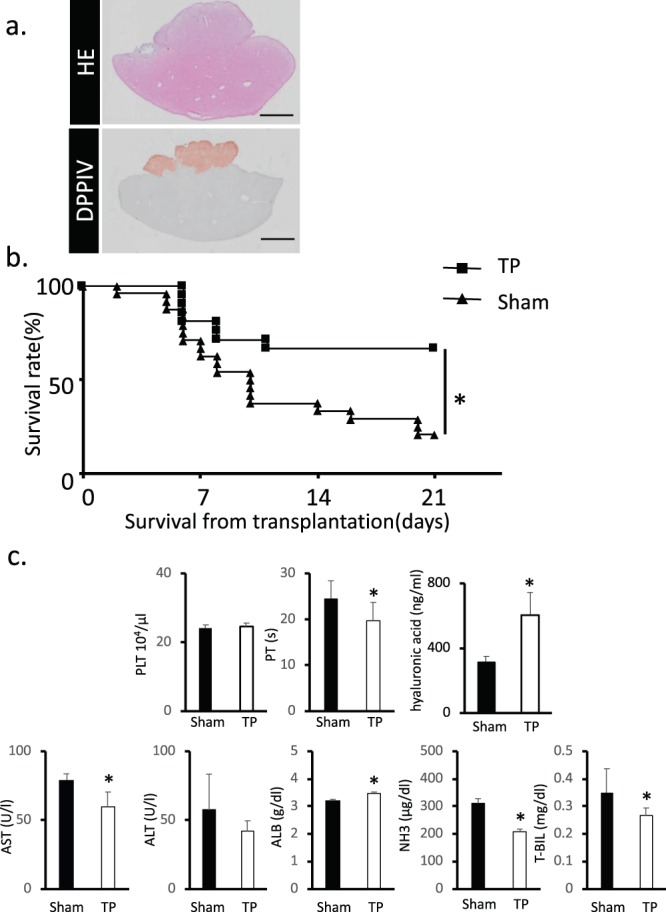
Figure 4.

Functional assessment of the transplanted fetal liver tissue in UPAL coating group. (a) Histological analysis of the fetal liver tissue that was engrafted on the surface of the middle lobe of the liver and covered by UPAL. DPPIV-stained living tissue is shown in red. Scar bar: 5 mm. (b) Measurement of the survival rate of transplanted fetal liver tissue in UPAL group. The survival rate in the transplanted group without UPAL serves as the control. Data are shown as mean ± SEM. *P < 0.01, log rank test, Sham: n = 24, TP: n = 21. (c) Levels of PLT, PT, hyaluronic acid, AST, ALT, T-BIL, NH3 and ALB were also comparable between the sham and transplanted groups after UPAL coating. Data are mean ± SE. *P < 0.05, vs. sham Mann-Whitney U test, n = 6–9.
