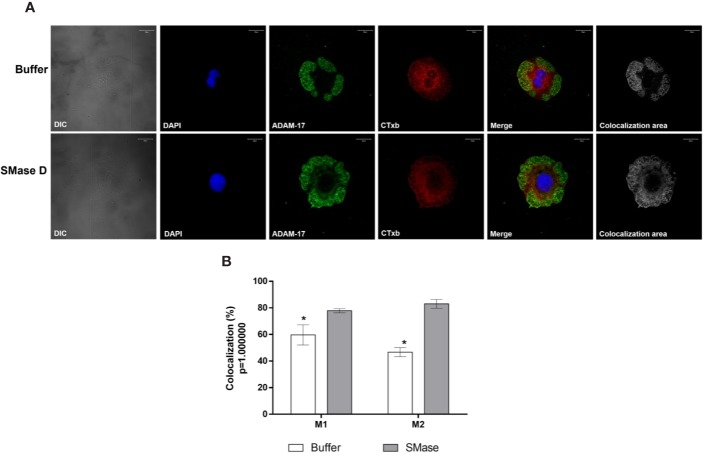
Figure 8.
Colocalization of ADAM-17 and Cholera Toxin subunit b in human keratinocytes, treated with SMase D. HaCaT cells were cultured on slides and treated for 2 h with buffer or SMase D (5µg/ml). Cells were stained with Moab anti-ADAM-17 (20 µg/ml), followed by RAM-FITC (1:50). GM1 containing lipid rafts were visualized, using the CTx-b/Alexa Fluor 555 and the nuclei counterstained with DAPI and slides were analyzed by CLSM. Scale bars represent 20 µm. (A) Colocalization of ADAM-17 and GM1 at the focal plane of 3.02 µm analyzed in cells treated with buffer or SMase D. Colocalization areas are shown as grayscale images. (B) Comparison between the colocalization of ADAM-17 and Cholera Toxin subunit b in cells treated with SMase D or buffer and represent means ± SEM from at least 10 images in two independent experiments and three different focal plans. Statistically analyzed by Two Way ANOVA followed by Tukey HSD test, using the GraphPad Prism 5.1. (*) Significant difference compared to SMase D (p < 0.05).

