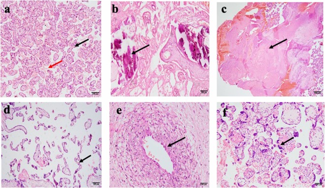
Figure 1.
Photomicrographs of placental changes observed among pregnant women from the study. (a) A non-PE placenta showing normal villi (black arrow) and intervillous spaces (red arrow). (b) Calcifications (black arrow) in a 33-week old PE placenta. (c) Infarction (coagulative necrosis) of large area of the placenta from ischaemia (black arrow) in 29-week placenta with intrauterine foetal death). (d) Accelerated villous maturation (thin finger-like or slender villi with reduced branching). (e) Atherosis showing accumulation of lipid laden macrophages within sub-endothelial area of arterial wall. (f) Increased syncytial knots showing densely stained and closely packed nuclei.

