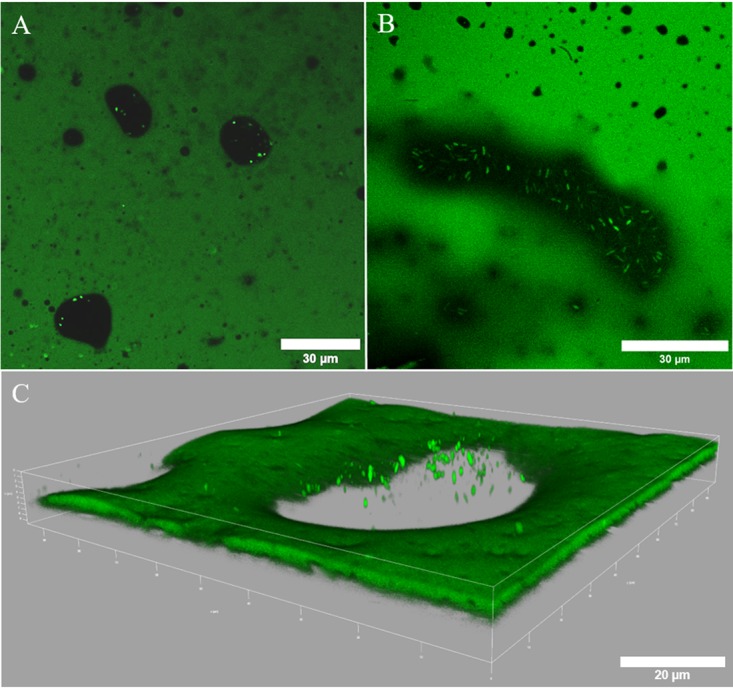
FIG 1.

CLSM fluorescence images of natural water droplets (black) dispersed in oil (green) from McKittrick (A, C) and La Brea (B) oil samples. Bright green dots represent microbial cells stained with Syto 9. (A, B) Two-dimensional view of different water droplets. (C) Three-dimensional view of different water droplets.
