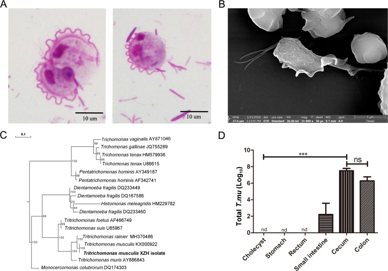
FIG 1.
Identification of a gut protozoan symbiont. (A) Representative microscopic image of the cecal content from protozoan-bearing mice, stained by Giemsa stain; scale bar, 10 μm. (B) Representative SEM image of purified protozoa. (C) Phylogenetic tree analysis according to the DNA sequences derived from the ITS rRNA region. The individual protozoal ITS sequence obtained from the NCBI GenBank database is indicated by its original species name followed by its accession number. The protozoan isolate that we found is in bold type. The evolutionary history was inferred using the RAxML method. Branch supports were computed out of 100 bootstrap trees. (D) The total number of T. musculis protozoa in the indicated tissues. The experiments were performed 2 times. The error bars represent standard deviations (n = 5 in each group). ***, P < 0.001; ns, no statistical significance; nd, not detected; T. mu, T. musculis.

