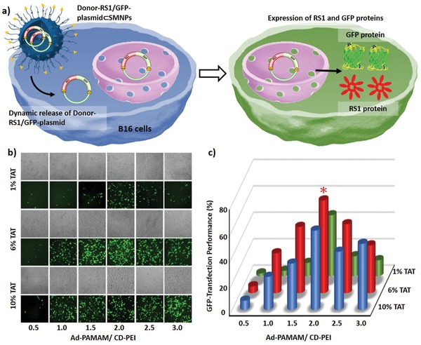
Figure 2.

a) Schematic illustration of green fluorescent protein (GFP) transfection in B16 cells treated by Donor‐RS1/GFP‐plasmid⊂SMNPs. b) Eighteen formulations of Donor‐RS1/GFP‐plasmid⊂SMNPs were prepared for the GFP‐transfection study, followed by fluorescence microscopy analysis. c) Quantitative analysis of the fluorescent micrographs revealed an optimal formulation (*) for Donor‐RS1/GFP‐plasmid⊂SMNPs.
