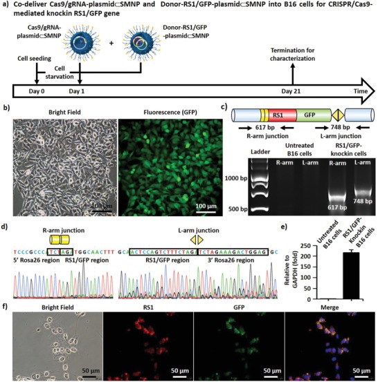
Figure 4.

a) A timeline depicting CRISPR/Cas9‐mediated knockin of RS1/GFP gene in growth‐synchronized B16 cells using both Cas9/sgRNA‐plasmid⊂SMNPs and Donor‐RS1/GFP‐plasmid⊂SMNPs. b) Bright‐field and fluorescence images of sorted RS1/GFP‐knockin B16 cells taken after 20 rounds of culture expansion. c) Two characteristic DNA fragments, i.e., the R‐arm junction (617 bp) and L‐arm junction (748 bp)—signifying the integration of RS1/GFP into the Rosa26 site—were detected by an electrophoretogram. d) Sanger sequencing was carried to test that the correct DNA sequences of the genome‐donor boundaries in the R‐arm and L‐arm junctions. e) RS1 gene expression levels observed by quantitative PCR. f) Representative immunofluorescence images of RS1/GFP‐knockin B16 cells.
